May 14, 2026
A huge congratulations to three outstanding PhD graduates from the Biomedical Optics Laboratory at the University of Houston! 🎓🎉
Jessica Gutierrez, Christian Zevallos Delgado, and Md Mobarak Karim have each successfully completed their doctoral degrees under the mentorship of Dr. Kirill Larin — and what an incredible achievement it is for all three!
Congratulations, Dr. Gutierrez, Dr. Zevallos Delgado, and Dr. Karim — the future is yours!

April 21, 2026
Congratulations to Md Mobarak Karim for successfully defending his PhD dissertation focused on the development of a multimodal imaging system for simultaneous structural and molecular imaging!

April 9, 2026
Congratulations to undergraduate Henry Chuo for presenting his work at Undergraduate Research Day. His research focused on assessing the structural and mechanical property changes in corneas in a model of keratoconus using second harmonic generation imaging and optical coherence elastography.

April 8, 2026
Congratulations to Jessica Gutierrez for successfully defending her PhD dissertation! The countless hours of research and hard work have led to this moment — welcome to the other side, Dr. Gutierrez!

March 25, 2026
Congratulations to Christian Zevallos-Delgado for successfully defending his PhD dissertation focused on mapping the mechanical properties of embryos with reverberant optical coherence elastography!

Feb 11, 2026
Congratulations to the lab for the newest publication: “Brillouin microscopic assessment of retinal stiffness during reactive Müller gliosis” in Experimental Eye Research.
March 1, 2026
Congratulations to the lab for the newest publication: “Quantitative comparison between optical coherence elastography, ultrasound-based shear wave elastography, and Brillouin imaging” in the Journal of Physics: Photonics.
Congratulations to the lab for a productive year, culminating in 16 peer-reviewed publications, including several high impact publications and reviews, over 20 conference proceedings, and hundreds of citations. The publications, presentations, successful experiments, and collaborative efforts each of you contributed to proved our technical capability, resilience, innovation, and commitment. As we move into the new year, let’s build on that momentum with even greater ambition, impact, and creativity.
- Aglyamov SR, Larin KV. Optical coherence tomography for noninvasive monitoring of drug delivery. Adv Drug Deliv Rev 2025;220:115571.
- Duvvuri C, Singh M, Lan G, Aglyamov SR, Larin KV, Twa MD. Determinants of Human Corneal Mechanical Wave Dispersion for In Vivo Optical Coherence Elastography. Transl Vis Sci Technol 2025;14:26.
- Chawla HS, Chen Y, Wu M, et al. Assessment of skin fibrosis in a murine model of systemic sclerosis with multifunctional optical coherence tomography. J Biomed Opt 2025;30:036007.
- Desai R, Tan F, Wu M, et al. Assessment of Skin in Patients With Systemic Sclerosis Using High-Frequency Ultrasound and Shear Wave Elastography: A Comparative Study With Histology, Molecular, and Clinical Parameters. Arthritis Care Res (Hoboken)
- Karim MM, Sun R, Khajavi B, et al. Multimodal optical coherence tomography and two-photon light sheet fluorescence microscopy for embryo imaging. J Biomed Opt 2025;30:060501.
- Karim MM, Nair A, Singh M, Hatami M, Aglyamov SR, Larin KV. Depth-Resolved Attenuation Coefficient Quantification During Murine Embryonic Brain Development. J Biophotonics 2025;18:e202500212.
- Nair A, Singh M, Aglyamov SR, Larin KV. Convolutional Neural Networks Enable Direct Strain Estimation in Quasistatic Optical Coherence Elastography. J Biophotonics 2025;18:e202400386.
- Nikitin PV, Chawla HS, Gutierrez J, et al. ALK5 inhibitor impact on bleomycin-induced systemic sclerosis mouse model via multifunctional optical coherence tomography. APL Bioeng 2025;9:046110.
- Mendieta M, Hatami M, Singh M, et al. Non-Invasive Measurement of Elasticity in Glioblastoma Multiforme Validates Decreased TMZ Sensitivity in Astrocyte Co-Culture. IEEE Open J Eng Med Biol 2025;6:287-295.
- Rohman L, Navia JC, Zevallos-Delgado C, et al. In Vivo Safety Evaluation of Acoustic Radiation Force for Optical Coherence Elastography of the Crystalline Lens. Transl Vis Sci Technol 2025;14:35.
- Schumacher J, Zevallos-Delgado C, Rohman L, et al. Clinical multimodal Brillouin microscopy-optical coherence elastography system for lens biomechanics. J Biomed Opt 2025;30:124511.
- Singh A, Singh M, Aglyamov SR, Mayerich D, Larin KV. Quantifying age and spatial variations of bone marrow elasticity with noncontact optical coherence elastography. J Biomed Opt 2025;30:124505.
- Singh A, Nair A, She Z, et al. Optical coherence elastography detects increased corneal stiffness in nonhuman primates with experimental glaucoma. J Biomed Opt 2025;30:124508.
- Singh A, Nair A, Saeidi Fard S, et al. Quantitative comparison between optical coherence elastography, ultrasound-based shear wave elastography, and brillouin imaging. J Phys Photonics
- Singh M, Hepburn MS, Kennedy BF, Larin KV. Optical coherence elastography. Nat Rev Meth Primers 2025;5:39.
- Zevallos-Delgado C, Mekonnen TT, Schill AW, Singh M, Aglyamov SR, Larin KV. Air-coupled ultrasound based noncontact reverberant optical coherence elastography. Light Adv Manuf 2025;6:1-8.
Jan 22, 2026
Congratulations to the lab for the newest publication: “Air-coupled ultrasound based noncontact reverberant optical coherence elastography” in Light: Advanced Manufacturing.
Jan 22, 2026
The Biomedical Optics Lab was excited to attend SPIE Photonics West 2026, one of the world’s leading conferences in optics and photonics!
It was a fantastic opportunity to engage with cutting-edge research, explore emerging technologies in biomedical imaging and photonics, and connect with researchers, industry leaders, and collaborators from around the globe.
We’re inspired by the innovative work showcased at the conference and look forward to bringing new ideas and collaborations back to our lab.
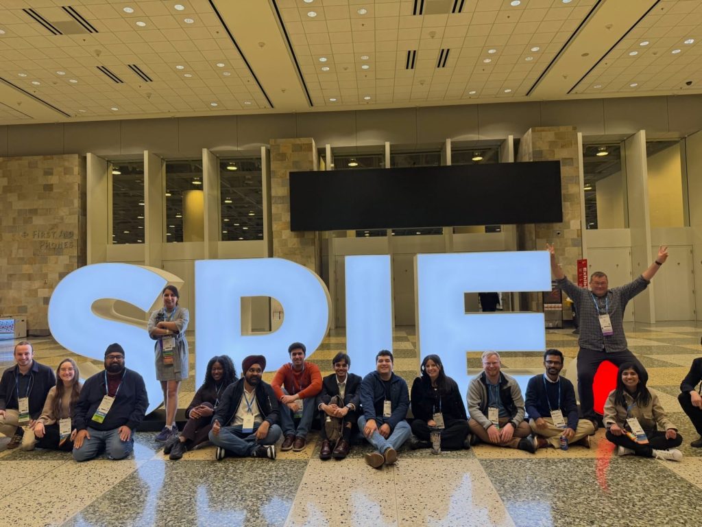
Photonics West 2026